A significant step in the bid to understand more about fusion divertor components has been reached after two different imaging methods were used by a team of scientists from Swansea University, Culham Centre for Fusion Energy, ITER in France, and the Max-Planck Institute of Plasma Physics in Germany.
As part of an initiative, they used both x-ray imaging and neutron imaging to try to decipher more about the robustness of parts.
The latter sees the sample placed in the path of a neutron beam. In addition to the radiation transmitted through the object being analysed (information that sheds light on the internal structure of the object), multiple images can be recorded as the sample rotates in the line of the beam. A similar approach using X-ray imaging can offer a higher resolution but cannot penetrate as much volume of material as neutron imaging – meaning both methods complemented one another. The neutron imaging work was carried out on the ISIS Neutron and Muon Source’s neutron imaging instrument, IMAT and X-ray imaging at Nikon Metrology, Tring.
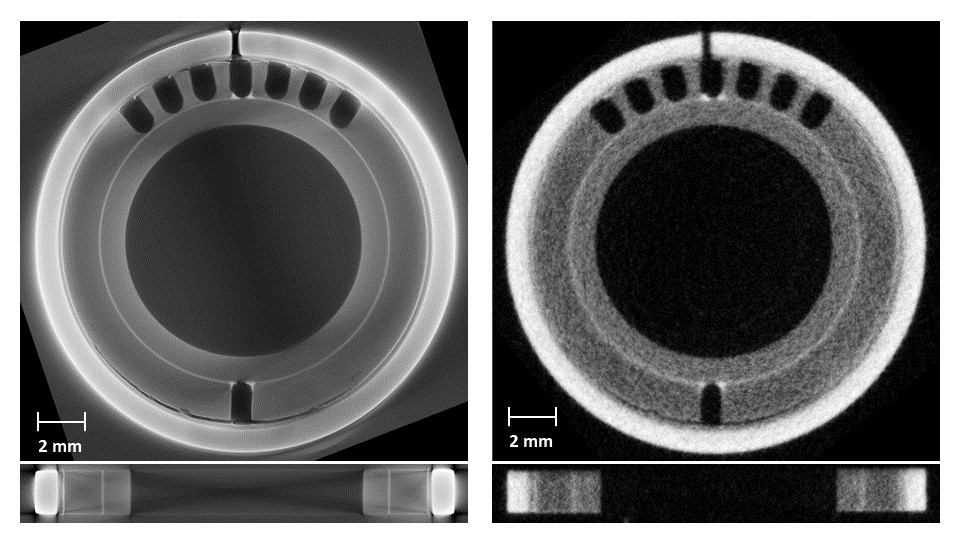
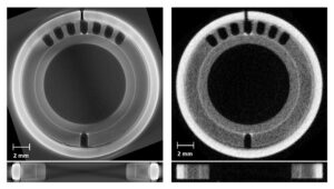
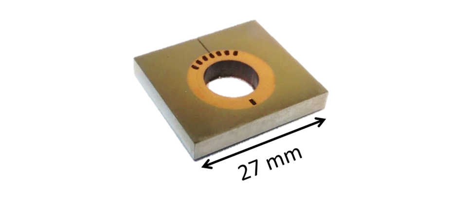
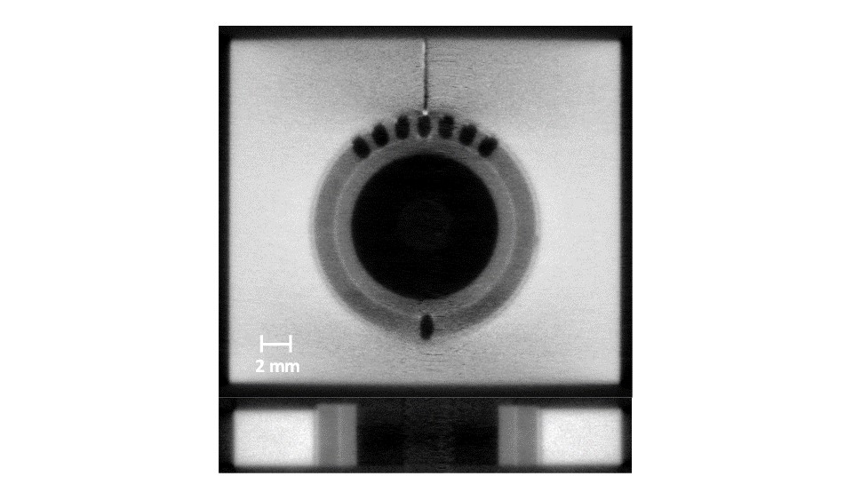
The research team focused on one critical component of a fusion energy device, called a monoblock, which is a pipe carrying coolant protected by armour. It was the first time a tungsten monoblock had been imaged by computerised tomography (a non-destructive technique for displaying a cross-section through a solid object using X-rays or alternative imaging). ITER will have around 320,000 monoblocks.
The divertor is seen as a vital component as it removes the heat and any impurities from the plasma. Because of its location and role in this, it is subject to an extreme heatload. The divertor components are cooled to counteract this, something done by pumping water coolant through the pipes.
Llion Evans, a researcher in Culham’s Technology Department and at Swansea University, said: “This work is a proof of concept that both these tomography methods can produce valuable data. In future, these complementary techniques can be used either for the research and development cycle of fusion component design or in quality assurance of manufacturing.
“The next step is to convert the 3D images produced by this powerful technique into engineering simulations with micro-scale resolution. This technique, known as image-based simulation (IBSim), enables the performance of each part to be assessed individually and account for minor deviations from design caused by manufacturing processes.”
Dr Triestino Minniti of the Science and Technology Facilities Council added: “Each technique had its own benefits and limitations. The advantage of neutron imaging over x-ray imaging is that neutrons are significantly more penetrating through tungsten. Thus, it is feasible to image samples containing larger volumes of tungsten. Neutron tomography also allows us to investigate the full monoblock non-destructively, removing the need to produce “region of interest” samples.”
Now that the team have demonstrated a whole tile can be imaged with neutron CT, the next step is to attempt this (again at IMAT) on a whole assembly of tiles that have been joined onto a coolant pipe.
Link to original article: https://doi.org/10.1016/j.fusengdes.2018.06.017
